Back Muscles Anatomy Reference : Back Muscles Anatomy Function Treatment - The muscles of the back can be arranged into 3 categories based on their location:
Back Muscles Anatomy Reference : Back Muscles Anatomy Function Treatment - The muscles of the back can be arranged into 3 categories based on their location:. These muscles stabilize the vertebral column and also have a role in proprioception and balance. Latissimus dorsi is a large muscle that, when atrophied, can cause significance asymmetry in the back. Muscles of the back can be divided into superficial, intermediate, and deep group. While it is a strong muscle for adduction, internal rotation and extension of the humerus, we can do without it. The former two groups, superficial and intermediate, are referred to as the extrinsic back muscles.
The tendon for latissimus dorsi can be harvested for tendon repairs or tendon transfers. These muscles stabilize the vertebral column and also have a role in proprioception and balance. Superficial back muscles, intermediate back muscles and intrinsic back muscles.the intrinsic muscles are named as such because their embryological development begins in the back, oppose to the superficial and intermediate back muscles which develop elsewhere and are therefore classed as extrinsic muscles. Attached to the posterior thorax. Axial section of the back showing the dorsal rami transmitting sensory neurons from the skin of the back to the spinal cord.
Back Muscles 28 Major Muscles Of The Back Earth S Lab from www.earthslab.com The basics of back pain and spinal anatomy. These muscles stabilize the vertebral column and also have a role in proprioception and balance. The deep back muscles, also called intrinsic or true back muscles, consist of four layers of muscles: Muscles of the back can be divided into superficial, intermediate, and deep group. The deep back muscles are posterior to the erector spinae. Back muscles the muscles of the back are a group of strong, paired muscles that lie on the posterior aspect of the trunk. The deep back muscles are posterior to the erector spinae. It extends from the external protuberance of the occipital bone to the lower thoracic vertebrae and laterally to the spine of the scapula.
The deep back muscles are posterior to the erector spinae.
We often don't think about their form and function, though, until they become a source of pain. The abdominal and back muscles maintain the spine's natural curves. The spaces between the vertebrae are maintained by intervertebral discs that act like shock absorbers throughout the spinal column to cushion the bones as the body moves. The deep back muscles, also called intrinsic or true back muscles, consist of four layers of muscles: Anatomy and biomechanics of the back muscles in the lumbar spine with reference to biomechanical modeling spine (phila pa 1976). They are short muscles associated with the spinous and transverse processes of the vertebrae. They provide movements of the spine , stability to the trunk, as well as the coordination between the movements of the limbs and trunk. Balance the weight of your head on top of your spine evenly distribute weights from your upper body into the lower extremities Back muscle diagram, back muscle diagram exercise, back muscle diagram pain, back muscles diagram a comprehensive view, back muscles diagram and ligaments, human muscles, back muscle diagram, back muscle diagram exercise, back muscle diagram pain, back muscles diagram a comprehensive view, back. The muscles of the back can be arranged into 3 categories based on their location: They are short muscles associated with the spinous and transverse processes of the vertebrae. Surface anatomy of the back showing bony landmarks on the left and cutaneous nerves on the right. The primary back muscles are divided into a total of five (5) systems, each system comprising.
Attached to the shoulder girdle. Balance the weight of your head on top of your spine evenly distribute weights from your upper body into the lower extremities They provide movements of the spine , stability to the trunk, as well as the coordination between the movements of the limbs and trunk. The reference for the following origin and insertion points is gray's anatomy (40 th edition) 1 unless otherwise referenced. It extends from the external protuberance of the occipital bone to the lower thoracic vertebrae and laterally to the spine of the scapula.
7 Essential Anatomy References Artstation Magazine from magazine.artstation.com On this page, you'll learn about each of these muscles, their locations and functional anatomy. Attached to the shoulder girdle. The primary back muscles are divided into a total of five (5) systems, each system comprising. Back, followed by 147 people on pinterest. Introduction the lower back (where most back pain occurs) includes the five vertebrae in the lumbar region and supports much of the weight of the upper body. Zygote scenes is a collection of scenes created by zygote media group with annotations identifying anatomical landmarks. The external abdominal oblique muscle lies on the sides and front of the abdomen and is the largest and the most superficial of the three flat muscles in this area. They are short muscles associated with the spinous and transverse processes of the vertebrae.
The trapezius has upper, middle, and lower groups of fibers.
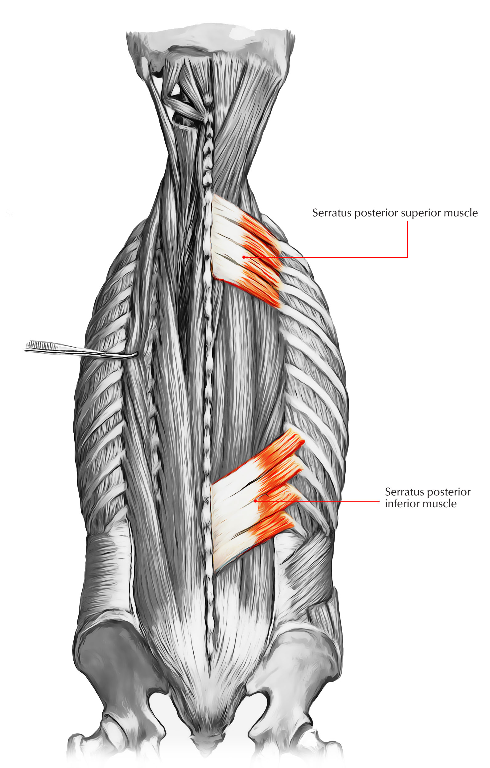
The reference for the following origin and insertion points is gray's anatomy (40 th edition) 1 unless otherwise referenced. The three deep muscles of the back include the semispinalis, multifidus, and rotatores. There are three major groups of back muscles: Anatomy and biomechanics of the back muscles in the lumbar spine with reference to biomechanical modeling spine (phila pa 1976). The deep back muscles are posterior to the erector spinae. Axial section of the back showing the dorsal rami transmitting sensory neurons from the skin of the back to the spinal cord. These muscles are divided into two major groups: The intrinsic back muscles are also referred to as primary back muscles.these muscles are also known as erector spinae (spinal erectors) or erector trunci (truncal erectors) since they specifically describe the primary function: Spinal anatomy is a remarkably intricate structure of strong bones, flexible ligaments and tendons, extensive muscles, and a highly sensitive spinal cord and nerve roots. The muscles of the back can be arranged into 3 categories based on their location: Muscles of the back can be divided into superficial, intermediate, and deep group. The latter group is the intrinsic muscle group. Zygote scenes is a collection of scenes created by zygote media group with annotations identifying anatomical landmarks.
Attached to the posterior thorax. All about the back muscles the back anatomy includes the latissimus dorsi, trapezius, erector spinae, rhomboid, and the teres major. The latter group is the intrinsic muscle group. Latissimus dorsi is a large muscle that, when atrophied, can cause significance asymmetry in the back. The deep back muscles, also called intrinsic or true back muscles, consist of four layers of muscles:
Male Muscle Drawing Reference from www.proko.com The human back, also called the dorsum, is the large posterior area of the human body, rising from the top of the buttocks to the back of the neck. See more ideas about anatomy, anatomy reference, anatomy drawing. 12 photos of the back muscle diagram & pain. Back muscle diagram, back muscle diagram exercise, back muscle diagram pain, back muscles diagram a comprehensive view, back muscles diagram and ligaments, human muscles, back muscle diagram, back muscle diagram exercise, back muscle diagram pain, back muscles diagram a comprehensive view, back. Anatomy and biomechanics of the back muscles in the lumbar spine with reference to biomechanical modeling spine (phila pa 1976). Back, followed by 147 people on pinterest. There are three major groups of back muscles: Attached to the shoulder girdle.
They provide movements of the spine , stability to the trunk, as well as the coordination between the movements of the limbs and trunk.
The human back, also called the dorsum, is the large posterior area of the human body, rising from the top of the buttocks to the back of the neck. While it is a strong muscle for adduction, internal rotation and extension of the humerus, we can do without it. The breadth of the back is created by the shoulders at the top and the pelvis at the bottom. They are short muscles associated with the spinous and transverse processes of the vertebrae. Muscles of the back can be divided into superficial, intermediate, and deep group. The back muscles are divided into two large groups: The trapezius has upper, middle, and lower groups of fibers. Within this group of back muscles you will find the latissimus dorsi, the trapezius, levator scapulae and the rhomboids. These muscles stabilize the vertebral column and also have a role in proprioception and balance. 12 photos of the back muscle diagram & pain. Surface anatomy of the back showing bony landmarks on the left and cutaneous nerves on the right. Back, followed by 147 people on pinterest. The latter group is the intrinsic muscle group.
These muscles stabilize the vertebral column and also have a role in proprioception and balance back muscles reference. This curve, called lordosis, helps to:


0 Komentar